
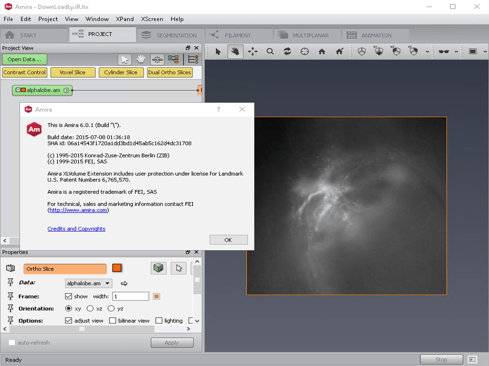
Download FEI Amira 6.0.1 – Advanced 3D and 4D Scientific Visualization and Analysis Software
FEI Amira 6.0.1 software is a sophisticated platform designed for advanced 3D and 4D scientific visualization and quantitative analysis of complex imaging data. Developed by TGS and later integrated into Thermo Fisher Scientific’s product portfolio, this scientific visualization software is tailored for professionals in biomedical research, life sciences, materials science, and medical imaging. It offers powerful tools to process, analyze, and understand multidimensional datasets derived from various imaging modalities.
Specialized 3D/4D Imaging for Biomedical and Materials Science
FEI Amira 6.0.1 excels in visualizing and analyzing multidimensional imaging data, making it an indispensable tool for fields such as biomedical research, neuroscience, cell biology, and materials science. The software effectively handles imaging data from sources including CT, MRI, confocal microscopy, and electron microscopy. Its capabilities allow researchers to delve into intricate structures and dynamics, supporting complex workflows for detailed visual analytics in specialized scientific domains.
Comprehensive Data Handling and Visualization Techniques
This scientific 3D visualization software supports the import of diverse imaging data types, including standard formats like DICOM, TIFF stacks, and LSM files, alongside proprietary formats from various microscopy and industrial CT systems. It provides high-quality volume and surface rendering options, enabling users to visualize cellular structures, anatomical regions, or material microstructures with realistic detail. Advanced multi-channel visualization, coupled with intuitive controls for lighting and transparency, allows for detailed examination of complex sample compositions and relationships.
Advanced Segmentation and Quantitative Analysis Modules
FEI Amira 6.0.1 offers a suite of advanced segmentation tools essential for biomedical image segmentation and quantitative analysis. Users can perform semi-automatic and manual segmentation using brushes, perform filament tracing for tubular structures, and conduct object counting for cellular or feature analysis. The software enables the extraction of precise statistical measurements, including volume, surface area, intensity distributions, and detailed analysis of network structures, providing quantitative data crucial for research publications and critical decision-making.
Spatial Graph and Network Analytics in Complex Datasets
A key capability of FEI Amira 6.0.1 is its specialized spatial graph analysis. This feature is particularly valuable for researchers studying complex networks within biological samples and materials, such as neuronal pathways, vascular systems, or porous material structures. The software can quantify intricate network characteristics, including path length calculations, branching pattern analysis, and detailed connectivity metrics, offering deep insights into the topology and function of these complex systems.
Enhanced Workflows and Automation with Python Integration
FEI Amira 6.0.1 includes robust registration and alignment tools designed to accurately fuse and analyze multi-modal and time-series imaging data. The platform significantly enhances custom automation workflows through its expanded Python scripting environment. This integration allows researchers to develop bespoke analytical pipelines, automate repetitive tasks, and connect with external machine learning libraries for advanced, data-driven image analysis, thereby streamlining complex research processes.
Performance Improvements and Updated Architecture in Version 6.0.1
Version 6.0.1 of FEI Amira introduces significant performance enhancements driven by its full native 64-bit architecture, enabling the seamless processing and visualization of exceptionally large datasets, including teravoxel-scale data. This updated architecture, combined with a faster rendering engine and other user interface improvements, ensures smoother interaction and higher efficiency. Furthermore, the release offers enhanced tools specifically for electron microscopy analysis, refining workflows for materials science and biological ultrastructure investigation with this powerful 64-bit visualization platform.
Frequently Asked Questions
What imaging data formats does FEI Amira 6.0.1 support?
FEI Amira 6.0.1 supports a wide range of medical and scientific imaging formats including DICOM, TIFF stacks, LSM microscopy files, industrial CT data, and 3D PDFs, enabling flexible data import from diverse imaging modalities.
How does FEI Amira enhance analysis of neuronal and vascular networks?
The software includes specialized spatial graph analysis modules that quantify complex network structures like neurons and blood vessels by measuring path lengths, branching, and connectivity, supporting advanced neuroscientific and biomedical research.
What improvements does version 6.0.1 bring for handling large datasets?
Version 6.0.1 introduces a full native 64-bit architecture, allowing the processing and visualization of very large datasets (teravoxel scale), combined with performance boosts like a faster rendering engine and improved user interface for smoother interaction with complex data.


Reviews
There are no reviews yet.